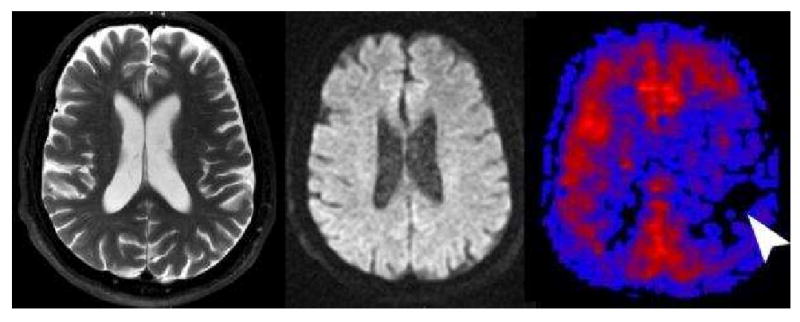
Figure 7. Label panels from Left to Right: A, B, C.

Tissue at Risk. This 70 year old male presented with transient ischemic attacks involving the right sided extremities. T2 (A) and diffusion (B) were normal except for mild diffuse cerebral atrophy. PASL CBF map (C) revealed significant hypoperfusion in the left frontal and parietal hemisphere. The patient had a follow-up CT Angiography showing a high grade stenosis of the left internal carotid artery that was surgically corrected without further clinical sequela.
