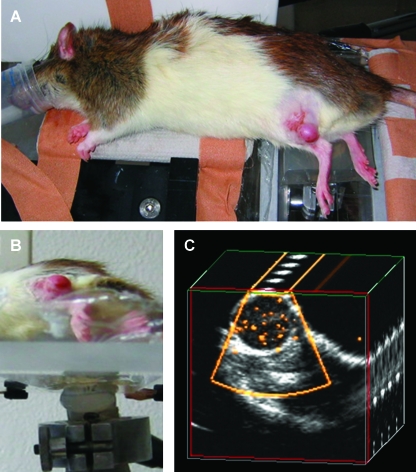
Figure 1.
SPAQ-based molecular imaging. (A) A rat with Dunning R3327-AT1 tumors implanted subcutaneously on both hind legs (white arrows) was placed on an ultrasound gel pad. (B) An ultrasound transducer was fixed under the navigable table, which can be moved in micrometer steps. The frame rate of the ultrasound Doppler system (25 Hz) was synchronized with the movement of the table (1.25 mm/sec), resulting in an incremental move of 50 µm between the consecutive ultrasound pulses. (C) During the ultrasound scan, the targeted microbubbles disintegrate and emit detectable signals (yellow dots within the tumor). The two-dimensional ultrasound images are reconstructed to a three-dimensional data set that is quantitatively analyzed by an automatic video densitometry system.

