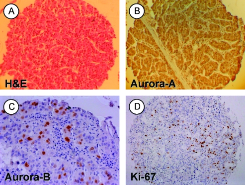
Figure 1.
Human HCC TMA. Representative samples of (A) an H&E.stained HCC tissue spot (original magnification, x100), (B) a HCC spot showing marked nuclear and cytoplasmic aurora-A protein localization (original magnification, x100), (C) a tumor spot with strong (++) expression of aurora-B (original magnification, x200), and (D) a respective tumor spot stained for the proliferation marker Ki-67 (MIB-1; original magnification, x100). Panels B to D were counterstained with hematoxylin.

