Figure 2.
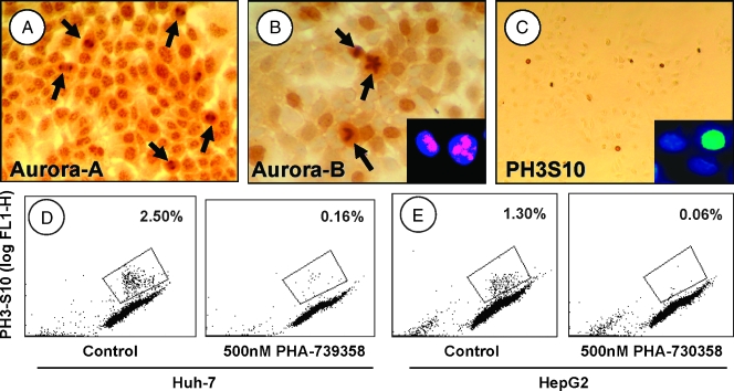
Aurora kinase expression in human HCC cells. (A) Huh-7 cells expressing aurora-A, which localizes to the centromeres or to the spindle pole (arrows; see text). (B) Aurora-B expression until the metaphase is seen as diffuse brown nuclear staining in Huh-7 (large panel) and HepG2 (insert, left cell) cells or in the spindle midzone during late mitosis (Huh-7, arrows; HepG2, insert, right cell). (C) Nuclear staining for phospho-H3S10 in Huh-7 and HepG2 (insert) cells quantified by flow cytometric analysis (D and E): Huh-7 (D) and HepG2 (E) cells, double stained with propidium iodide (DNA content) and anti-phospho-H3S10. Left panels show controls; right panels show the respective cells treated with 500 nM PHA-739358. The gates indicate phospho-H3S10.positive cells. Original magnifications: x200 (A), x400 (B), x100 (C), x600 (inserts).

