Figure 3.
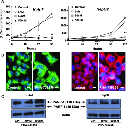
Antiproliferative effects of PHA-739358 treatment in vitro. Left panels, Huh-7; right panels, HepG2. (A) Antiproliferative effect of PHA-739358 in rapidly proliferating Huh-7 cells (analyzed after 96 hours) and moderately proliferating HepG2 cells (for up to 120 hours). Significant antiproliferative effects of PHA-739358 at 50 and 500 nM compared with DMSO controls. *P < .05; **P < .01 of 50 nM/500 nM PHA-739358 compared with controls. (B) Immunohistochemistry for cytokeratin 18 as a cytoplasmic marker of HCC cells treated with DMSO control or 50 nM PHA-739358. Treatment with the compound for 48 hours resulted in large nuclei in Huh-7 (green) and HepG2 cells (red) indicating endoreduplication. Original magnification, x400; scale bars, 10 µm; counterstained with Hoechst-33258. (C) Effects of PHA-739358 treatment for 24 hours on PARP-1 cleavage as an indicator of caspase mediated apoptosis. The 116-kDa band indicates full-length PARP-1; the 89-kDa band represents the large fragment of PARP-1, which results only in PHA-739358-treated Huh-7 but not in HepG2 cells.

