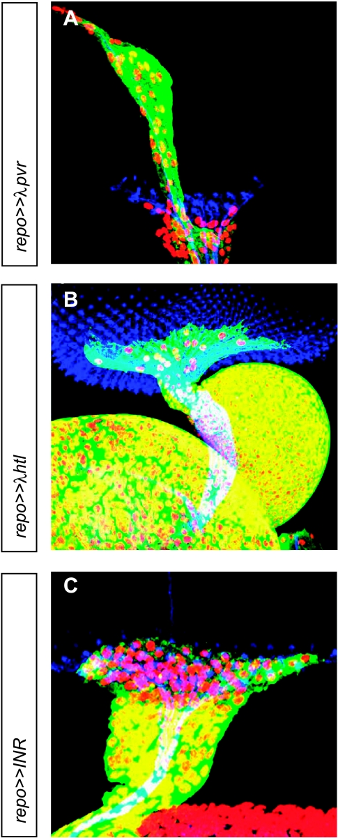
Figure 4.
Eye imaginal discs of third instar larvae with overexpression of other tyrosine kinase receptors in glial cells. Immunofluorescence staining of (A–C) eye imaginal discs. Nuclei of glial cells are red (α-Repo), glial cytoplasms are green (α-GFP), and photoreceptor neurons are blue (α-HRP). Images are projections of confocal image stacks. (A) Ectopic gene expression of activated pvr, the Drosophila homolog of PDGFR/VEGFR, resulted in overmigration of glial cells along BN. (B and C) Ectopic gene expression of λhtl (homolog of activated FGFR1) and INR (insulin receptor) resulted in a thickened optic stalk due to increased numbers of glial cells.

