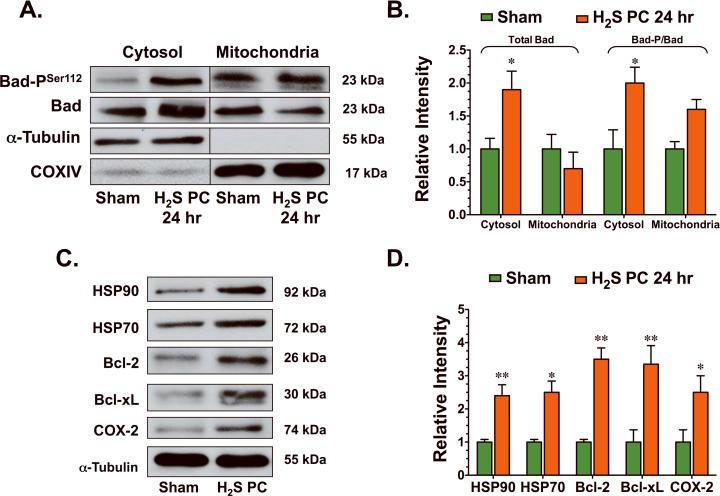
Figure 6.
H2S increased the expression of anti-apoptogens. (A–B) Representative immunoblots and densitometric analysis of phosphorylated Bad at serine residue 112 (Bad-PSer112) and total Bad (cytosolic and mitochondrial fractions) and (C–D) HSP90, HSP70, Bcl-2, Bcl-xL, and COX-2 24 hr following the administration of H2S. Values are means ± S.E.M. for an n of 4–5 animals for each group. *p<0.05, **p<0.01 vs. Sham.

