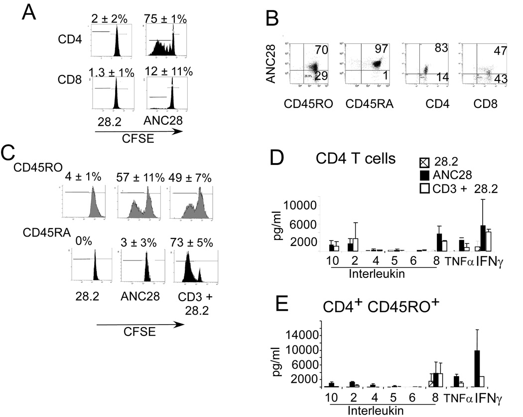
Figure 2. Selective expansion of CD4CD45RO cells by ANC28.
CFSE-labeled purified (A) CD4, CD8 cells stimulated with 28.2 or ANC28 and (C) CD4CD45RO, CD4CD45RA stimulated with ANC28, 28.2 or CD3+28.2 were analyzed by flow cytometry on day 6. Mean percent of dividing cells ± SD, p<0.05 ANC28 vs. 28.2 and p<0.05 CD4ANC28 vs CD8ANC28, N = 3 different donors. Expansion of CD45RO was comparable with ANC28 or CD3+28.2 stimulation and the CD4CD45RA subset of T cells was unresponsive to ANC28. (B) Purified CD4, CD8, CD45RA or CD45RO were stained with PE–conjugated ANC28 and analyzed by flow cytometry for CD28 expression. Low percentages of CD28+cells were present in CD8+ sub-population (N=2).
Mean ± SD indicate the production levels of indicated cytokines from (D) CD4 or (E) CD4CD45RO cells. Culture supernatants two days after of stimulation by ANC28, 28.2 and 28.2+CD3 were assayed by cytokine bead arrays. ANC28 but not 28.2 induced high levels of inflammatory cytokines including IL-2, IL-8, and TNF-α and IFN-γ. N = 3 different donors. (E) Purified CD4, CD8, CD45RA or CD45RO were stained with PE–conjugated ANC28 and analyzed by flow cytometry for CD28 expression. Low percentages of CD28+cells were present in CD8+ sub-population (N=2).

