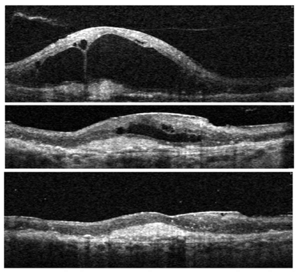
FIGURE 4.

Exudative AMD with VMT, surgical outcome (Patient 4). Vertical Spectral OCT scans through the center of the macula are shown. (Top) Baseline scan shows vitreous adherent to the apex of large cavities within edematous retina, and an underlying CNV complex; this fluid persisted despite eight previous monthly injections of intravitreal Bevacizumab. (Middle) Image shows center of macula one month after vitrectomy with removal of the attached hyaloid membrane. Fluid is reabsorbing (note: the scale is adjusted to facilitate direct comparison with the other scans). (Bottom) (same magnification as top) six months follow-up: complete reabsorption of intra- and sub-retinal fluid. Monthly Bevacizumab treatment was continued after surgery. Vision improved by one line, from 20/400 to 20/320.
