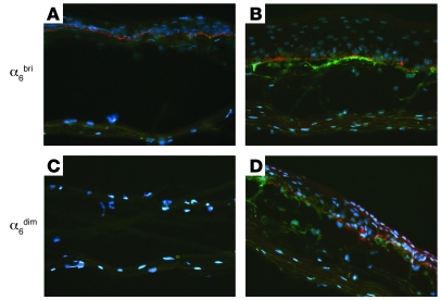
Figure 5. Increased deposition of LAMA5 by α6dim keratinocytes cocultured with HD-1bri pericytes in OCs.
Immunofluorescence staining for LAMA5 (mAb 4C7, red) and HD-1 antigen (green) performed on 3-μm frozen sections of OCs, showing epithelial sheets regenerated by α6bri (combined stem and TA; A and B) or α6dim early differentiating keratinocytes (C and D) seeded on a dermal equivalent containing either P7 HFFs alone (A and C) or P7 HFFs plus HD-1bri pericytes (B and D). Nuclei were counterstained with DAPI (blue). LAMA5 immunostaining in the epidermal-dermal junction was observed in all OCs reconstituted with α6bri keratinocytes, irrespective of the presence of pericytes (A and B). In contrast, OCs reconstituted with α6dim keratinocytes cocultured with HD-1bri pericytes (D) displayed largely increased LAMA5 levels compared with controls (C). HD-1 staining revealed the localization of pericytes in close proximity to the epidermal cells in OCs, where they were added back to the dermal equivalents (B and D). Original magnification, ×20.

