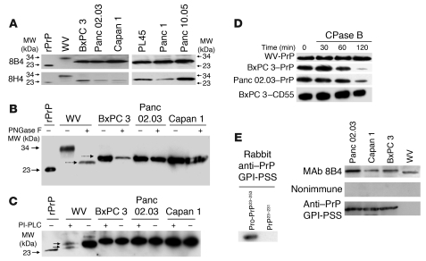
Figure 2. PrP in the PDAC cell lines exists as pro-PrP.
(A) Immunoblots show PrP from WV cells has a MW of 34 kDa, while PrP from the PDAC cell lines has a MW of 26 kDa. A recombinant PrP (rPrP) produced in E. coli is included as a control and MW marker. (B) Immunoblots show treatment of PrP from WV cells with endoglycosidase-F (PNGase F) reduces its MW from 34 kDa to 25.5 kDa. But identical treatment does not change the mobility of PrP from the PDAC cell lines. Deglycosylated PrP from WV cells migrated slightly faster than PrP from the PDAC cell lines (dashed arrows). (C) Immunoblots show PrP from WV cells is sensitive to PI-PLC, as shown by the appearance of a smaller PrP species (bottom arrow) in addition to the PNGase F–treated species (top arrow), but PrP from the PDAC cell lines is resistant to PI-PLC. (D) Immunoblots show that while PrP from the 2 PDAC cell lines is sensitive to CPase B, PrP from WV cells is resistant. CD55 from BxPC 3 cells is also resistant to CPase B. (E) Immunoblots show a rabbit antiserum specific for the PrP GPI-PSS reacts with recombinant pro-PrP (rPro-PrP23–253) but not with recombinant mature PrP (rPrP23–231). The anti–GPI-PSS antiserum also reacts with pro-PrP from the PDAC cell lines but does not react with the PrP from WV cells.

