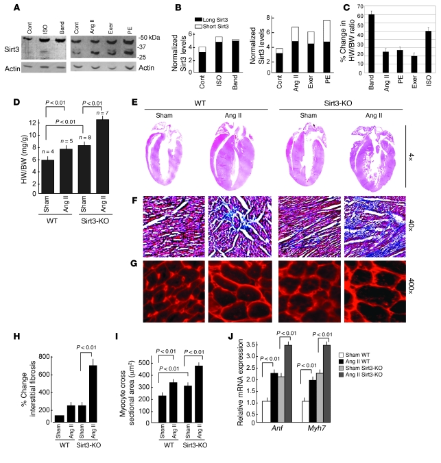
Figure 1. Sirt3 is required to block cardiac hypertrophic response.
(A) Expression levels of 2 forms of Sirt3 in different models of cardiac hypertrophy. Western blotting analysis of heart samples of mice subjected to chronic infusion of agonists, ISO, Ang II, or PE as well as of aortic banding (Band) or forced swimming exercise (exer). (B) Quantification of 2 forms of Sirt3 in different models of hypertrophy. (C) Percentage of cardiac hypertrophy in response to different stimuli. Values are mean ± SEM, n = 5–10. (D) HW/BW ratios of WT and Sirt3-KO mice infused with either vehicle (sham) or Ang II for 14 days. Values are mean ± SEM. (E) H&E-stained sections of hearts from WT and Sirt3-KO mice subjected to Ang II–mediated hypertrophy show gross changes of cardiac hypertrophy. (F) Sections of hearts stained with Masson’s trichrome to detect fibrosis (blue). (G) Heart sections stained with wheat germ agglutinin to demarcate cell boundaries. Original magnification, ×4 (E); ×40 (F); ×400 (G). (H and I) Quantification of fibrosis and myocyte cross-sectional area in control (sham) and Ang II–treated WT and Sirt3-KO mice hearts. (J) Anf and Myh7 mRNA levels in heart samples of control (sham) and Ang II–treated WT and Sirt3-KO mice. Mean ± SEM (n = 4–8). Cont, control.

