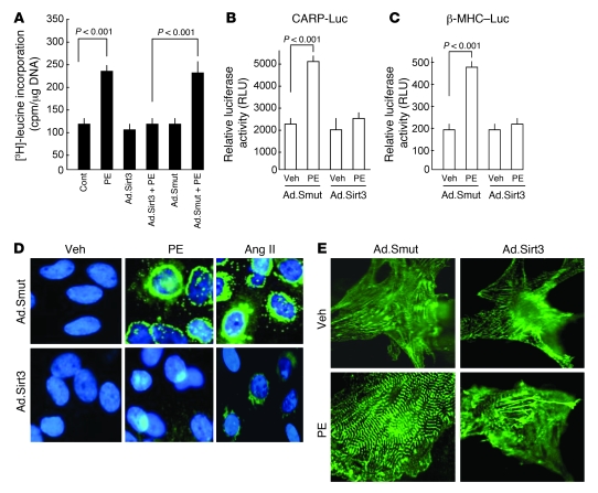
Figure 2. Sirt3 overexpression blocks cardiac hypertrophic response in vitro.
(A) Rat cardiomyocytes were overexpressed with Sirt3 WT (Ad.Sirt3) or mutant virus (Ad.Smut) and then treated with PE (20 μM) for 48 hours. Incorporation of [3H]-leucine into total cellular protein was determined and normalized to DNA content of the cells. Values are mean ± SEM (n = 5). (B and C) Cardiomyocytes expressing Ad.Sirt3 or Ad.Smut viruses were transfected with a CARP promoter/luciferase reporter vector (CARP-Luc) or β-MHC promoter/luciferase reporter vector (β-MHC–Luc). Cells were treated with vehicle (Veh) or PE (20 μM), and the luciferase activity was measured 48 hours after treatment. A β-gal/reporter plasmid was used as a reference control. Values are mean ± SEM (n = 3). (D) Cardiomyocytes were infected with the indicated adenoviruses and then stimulated with PE (20 μM), Ang II (2 μM), or vehicle for 48 hours. ANF release (green) was determined by staining cells with anti-ANF antibody. DAPI stain was used to mark the position of nuclei. (E) Reorganization of sarcomeres after PE treatment of cells. Cells were treated as in D and immunostained with α-actinin antibody for visualization of sarcomeres. Original magnification, ×630 (D); ×1,000 (E).

