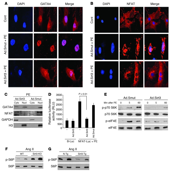
Figure 5. Sirt3 inhibits activation of transcription and translation regulators involved in development of hypertrophy.
(A and B) Rat cardiomyocytes were infected with Ad.Sirt3 or Ad.Smut viruses and then stimulated with either vehicle or PE (20 μM) for 2 hours. Cells were stained for GATA4 (A) or NFAT (B), and subcellular localization of factors was determined by confocal microscopy. Positions of nuclei were determined by DAPI stain (blue). Original magnification, ×630 (A and B). (C) Cardiomyocytes were infected with viruses and treated with PE as in A. Cytoplasmic (Cyto) and nuclear (Nucl) fractions of myocytes were generated and analyzed by Western blotting. (D) Cardiomyocytes expressing the indicated adenoviruses were transfected with a basic luciferase plasmid (B-Luc) or NFAT-responsive/luciferase (NFAT-Luc) reporter plasmid. On the second day after transfection, cells were treated with PE (20 μM), and the luciferase activity was determined 24 hours after transfection. Values are normalized with the protein content of the cell (mean ± SEM, n = 5). (E) Cardiomyocytes were infected with the indicated adenoviruses and induced with PE (20 μM). Cells were harvested at different time points after PE treatment, and the lysate was analyzed by Western blotting. (F and G) Heart extracts of mice subjected to Ang II–mediated hypertrophy were analyzed by Western blotting. Results are shown for 2 mice of the same group. WT and N-Tg mice are controls of the same genetic background for Sirt3-KO and Sirt3-Tg mice, respectively.

