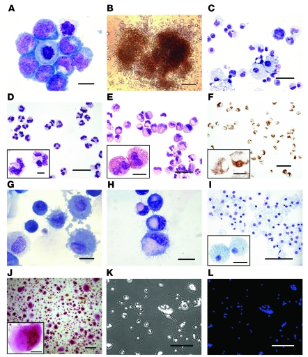
Figure 3. Morphology and cytochemical features of H1 hESC–derived myeloid progenitors and differentiated myelomonocytic cells.
(A) Wright-stained cytospins of H1-derived CD235a/CD41a–CD45+ cells after 2 days expansion with GM-CSF (scale bar: 10 μm). (B) Typical GEMM-CFC generated from day 2 GM-CSF–expanded cells (scale bar: 200 μm). Wright-stained cytospins of GEMM-CFC (scale bar: 40 μm) (C), hESC-derived mature neutrophils (scale bar: 25 μm; inset, 5 μm) (D), eosinophils (scale bar: 40 μm; inset, 10 μm) (E), CD1a+ cells isolated from DC (scale bar: 10 μm) (G) and LC (scale bar: 10 μm) (H) cultures, and macrophages (scale bar: 100 μm; inset, 20 μm) (I). (F) Cytochemical staining for eosinophil-specific (cyanide-resistant) peroxidase of hESC-derived eosinophils (scale bar: 50 μm; inset, 10 μm). (J) Cytochemical staining for TRAP of hESC-derived osteoclasts (scale bar: 300 μm; inset, 100 μm). Phase-contrast (K) and corresponding DAPI staining (L) of osteoclasts to demonstrate multinucleation (scale bar: 200 μm).

