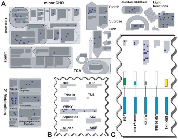
Figure 4.
Overview of genes regulated in pathogen associated contrasts. The gray areas inside the individual diagrams of the functional categories represent all genes present on the ATH1 chip. Dark blue squares highlight genes regulated in contrasts of the “pathogen” cluster. A) Regulation of cell wall genes (upper left), alkaloids which fall into the category “N-misc.” of “secondary metabolism” and “Light Reactions” of photosynthesis (upper right) is apparent. B) Part of the “transcription” map indicating regulation of WRKY transcription factors. C) Section of the “receptor like kinases” map indicating regulation of DUF26 kinases. Figure reading example: In subfigure C, a total of 41 DUF26 kinases are represented on the ATH1 chip of which 9 are regulated after pathogen exposure.
The figure is based on maps from the pathway analysis program MapMan (Usadel et al. 2005).

