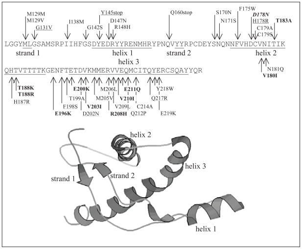
Figure 1.
Sequence and mutations in the C-terminal domain of huPrP together with the ribbon drawing of the corresponding 3D structure (positions 125 to 228 of 1HJM). The secondary structure segments are denoted by underscores. Bold indicates pathogenic mutations associated with the CJD phenotype, underline indicates GSS, and italic indicates FFI.

