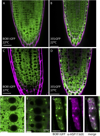
Figure 4.
Temperature-dependent BOB1 subcellular localization. Both BOB1:GFP (A and E) and 35S:GFP (B) are distributed throughout the cytoplasm of root cells at 22°C. BOB1:GFP forms granules at 37°C (C and F), while 35S:GFP localization at 37°C is unchanged (D). BOB1:GFP (G) and an α-HSP17.6 antibody (H) were used to show that these proteins colocalize in HSGs (white in I). Cell walls stained with propidium iodide are shown in magenta in A to F. Bars = 25 μm in A to D and 5 μm in E to I.

