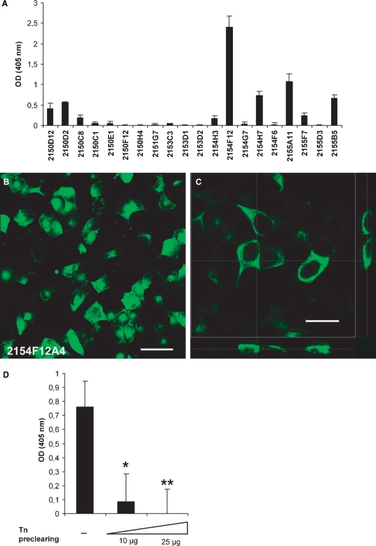
Fig. 3.
Selection of specific anti-Tn antibodies. (A) Mean values ± SD of optical density (OD) obtained from an ELISA screening of anti-Tn clones on MCF7 cells grown on a 96-well plate. (B) Representative immunofluorescence analysis performed on MCF7 cells grown on a glass coverslip. The green staining indicates cell surface binding of mAb 2154F12A4. Bar: 50 μm. (C) A higher magnification image of MCF7 cells stained with mAb 2154F12A4 and its xz (lower panel) and yz (right panel) sections evidenced the binding of mAb to the cell membrane. Bar: 25 μm. (D) Inhibition of mAb 2154F12A4 binding to the surface of MCF7 cells after solid-phase preclearing with Tn. The values report the mean values ± SD of three ELISA assays performed in duplicate, indicating the reactivity of the mAb on a monolayer of MCF7 cells after pre-incubation with scalar doses of Tn (10 and 25 μg) adsorbed onto a plastic microtiter plate. *P = 0.006; **P = 0.003.

