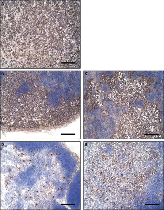
Fig. 5.
Cytokeratin staining of MCF7-derived tumors and mouse lymph nodes. (A–E) Immunoperoxidase staining of mouse cryostat sections. (A) MCF7 flank tumor; (B and C) right inguinal and axillary lymph nodes; (D and E) left inguinal and axillary lymph nodes. Brown staining indicates the pan-cytokeratin positive cells, a marker for human carcinoma cells. Notice the large metastatic nests in the right lymph nodes, ipsolateral to the flank tumor. Nuclei are evidenced in blue by hemallume staining. Bars: 120 μm.

