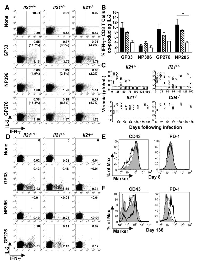
Fig. 2.
Severe CD8+ T cell exhaustion and viral persistence in the absence of IL-21. Splenic CD8+ T cell responses and viral titers were evaluated following LCMV-Cl 13 infection of Il21+/+, +/−, and −/− mice. (A) Flow cytometric analysis of intracellular cytokine staining for IFN-γ and IL-2 production by CD8+ T cells at eight days following infection after restimulation without or with the indicated peptide epitopes. Gated CD8+ T cells are shown and the percentages of CD8+, IFN-γ+ cells that co-produce IL-2 are reported in parentheses. (B) Percentages of epitope-specific CD8+, IFN-γ+ cells that coproduce IL-2 at eight days following infection. Error bars are SEM; * P<0.05 by comparison with Il21+/+ group. (C) Serum viral titers over time following LCMV-Cl 13 infection of Il21+/+, +/−, −/−, and Cd4−/− mice. Results from individual mice are shown; the dotted line represents the limit of detection. (D) IFN-γ and IL-2 production by LCMV-specific CD8+ T cells at 136 days following infection. Gated CD8+ T cells are shown. (E and F) CD43 and PD-1 expression by GP33 tetramer+ CD8+ T cells from Il21+/+ (shaded), +/− (dashed line), and −/− (bold line) mice at eight (E) and 136 days (F) post-infection. The Il21+/− data shown in (D) and (F) are from mice that were aviremic at the time of analysis. Representative or composite data are shown from two independent experiments (n=3–6).

