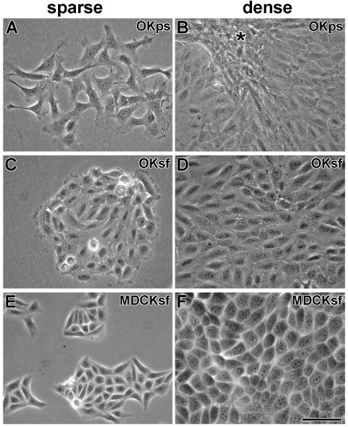
Figure 1. Morphology of OK and MDCK cells in complete medium with or without streptomycin.

A). At low density, OK cells raised in penicillin-streptomycin culture media (OKps) look fibroblastic. B) At high density, OKps cells retain a fibroblastic morphology and crowd together (*). C) At low density, OK cells, grown in streptomycin-free media (OKsf) for at least 7 weeks, cluster together and have an epitheloid appearance. D) At high density, confluent OKsf cells retain their epitheloid appearance. E) At low density, MDCK cells, grown in streptomycin-free media for at least 7 weeks (MDCKsf), cluster together and appear epitheloid (as in penicillin-streptomycin media). F) At high density, confluent MDCKsf cells retain their epitheloid appearance. Scale bar = 50 μm.
