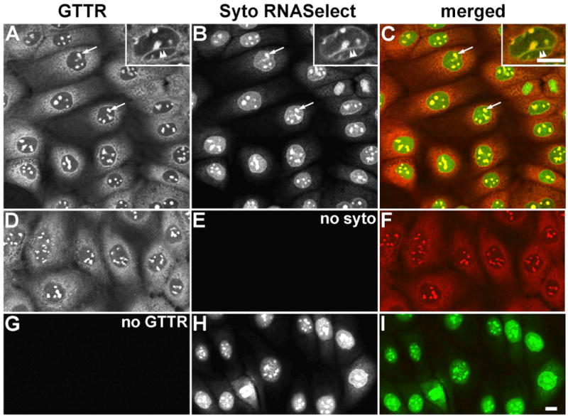
Figure 4. Distribution of GTTR in methanol-fixed MDCK cells double-labeled with Syto RNASelect.

A) GTTR is diffusely distributed throughout the cytoplasm, and strongly labels the intra-nuclear structures (arrows), and trans-nuclear tubules (double arrowhead in inset). B) Syto RNASelect strongly labels the globular intra-nuclear structures (arrows), and trans-nuclear tubules (double arrowhead in inset). C) Merged images of (A) and (B), show co-localization of both GTTR and SYTO RNASelect fluorophores as yellow in globular intra-nuclear structures (arrows), and trans-nuclear tubules (double arrowhead in inset). D) Fluorescent GTTR-loaded cells. E) Cells in (D), not treated with Syto RNASelect, display negligible non-specific 515 nm fluorescence. F) Merged image of (D) and (E). G) Cells treated with SytoRNA Select only (in H) display no bleed-through fluorescence in red (GTTR) channel. H) Cells treated with Syto RNASelect. Note mitotic figure lower left. I) Merged image of (G) and (H). Scale bars = 10 μm.
