
Figure 5.
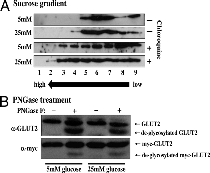
Intracellular GLUT2 distribution and glycosylation. A, Min6B1 cells were incubated for 24 h with 5 or 25 mm glucose (Glu) in the presence or absence of 50 μm chloroquine (Chloro). The cells were homogenized and subjected to sucrose gradient centrifugation as described in Materials and Methods. The gradient fractions were then extracts with Laemmli sample buffer and immunoblotted for the presence of the endogenous GLUT2 protein. B, Min6B1 cells were transfected with 2 μg of Myc-GLUT2 plasmid and incubated 24 h with 5 or 25 mm glucose. Cell extracts were then prepared and treated with and without PGNase F. The samples were then subjected to SDS-PAGE and immunoblotted for the endogenous GLUT2 protein or the exogenous Myc-GLUT2 protein. PNGase, N-glycopeptidase.