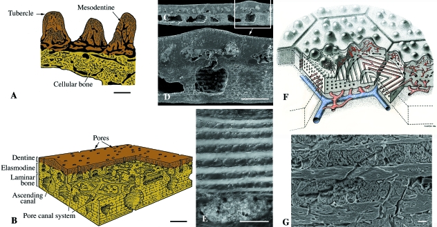
Fig. 8.
Osteostraci (Silurian). Schematic illustrations (A,B,F) and SEM sections (C–E,G) of osteostracan integumentary elements. (A) Procephalaspis. The basal plate of cellular bone is covered by a layer of tubercles composed of mesodentine, and a thin layer of enameloid. (B) Diagram illustrating the structure of cosmine-like tissue (including the pore-canal system) of the integumentary skeleton of Tremataspis. (C,D) Scale-like element from an unidentified thyestiid. (E) Detail demonstrating the plywood-like tissue (putative elasmodine) of the middle layer of a Tremataspis scale. (F) Reconstruction of the inter-relationships between the polygonal plate-like tesserae of the head-shield and the underlying, richly vascularized region from the osteostracan Alaspis rosamundae (from an unpublished manuscript by Tor Ørvig). Two series of canals are figured in blue and red. (G) Close-up of putative elasmodine in the integumentary skeleton of Hemicyclaspis. Scale bars: A–D: 150 µm; E: 60 µm; G = 10 µm.

