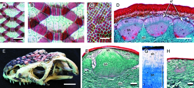
Fig. 5.
Lepidosaur osteoderms. (A–C) Alizarin red single-staining. (D,F–H) Longitudinal sections (dorsal towards the top). (A,B) Egernia sp. (Scincidae, Extant). Among scincids, most postcranial osteoderms overlap one another and demonstrate a compound or fractured morphology. (C) Tarentola mauritanica (Gekkota, Extant) postcranial osteoderms with a granular morphology. (D) Postcranial osteoderms from T. annularis (Gekkota, Extant) stained with Masson's trichrome. Each osteoderm has two distinct tissue regions. The superficial region resides entirely within the stratum superficiale, and is collagen-poor with virtually no incorporated cells. The basal region resides within the stratum compactum, and consists of compact (cellular) bone. Sharpey's fibres anchor both regions within the surrounding dermis. (E–H) Heloderma horridum (Helodermatidae, Extant). (E) Adult skull demonstrating the presence of osteoderms. Osteoderms from the left lateral surface have been removed to reveal the underlying cranial elements. Heloderma horridum osteoderms stained with Masson's trichrome (F,H) and toluidine blue (G). Similar to Tarentolaspp., H. horridum skeletally mature osteoderms (F,G) have a superficial collagen-poor region and a basal region composed of compact bone. (H) Skeletally immature osteoderm (white asterisk) demonstrating the earliest stages of mineralization. Note the absence of an osteoblast-rich condensation. This mode of ossification is consistent with bone metaplasia. The superficial region develops later during ontogeny. bh (basal region of bone-rich tissue), sf (Sharpey's fibres), sh (superficial region of unidentified skeletal tissue), stc (stratum compactum of the dermis), sts (stratum superficiale of the dermis). Scale bars: A = 1 mm, C = 0.5 mm, D = 50 µm, E = 20 mm, F–H = 100 µm.

