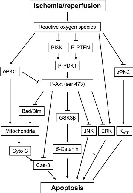
Figure 2.
The diagram shows the cell signaling pathways, which were involved in postconditioning. Reperfusion after stroke leads to ROS eruption, which causes dysfunction of the Akt cell signaling pathway, increases δ PKC activity, although decreases ε PKC activity; in addition, ROS also activates JNK and ERK activity. Furthermore, the Akt pathway is associated with the JNK and ERK pathways. The PI3K–Akt inhibition directly results in dephosphorylation of GSK3β and β-catenin degradation, and indirectly causes cytochrome c release and caspase-s activity. ε PKC may activate KATP channel resulting in neuroprotection. ROS, reactive oxygen species; Cyto C, cytochrome c; Cas-3, caspase-3; GSK 3 β, glycogen synthase kinase 3β; PI3K, phosphoinositide 3-kinase; PKC, protein kinase C; P-Akt, phosphorylated Akt; P-PTEN, phosphorylated phosphatase and tensin homolog deleted on chromosome 10; P-PDK1, phosphorylated phosphoinositide-dependent protein kinase-1; JNK, c-Jun N-terminal kinases; ERK, extracellular signal-regulated kinases; KATP channels, ATP-sensitive potassium channels.

