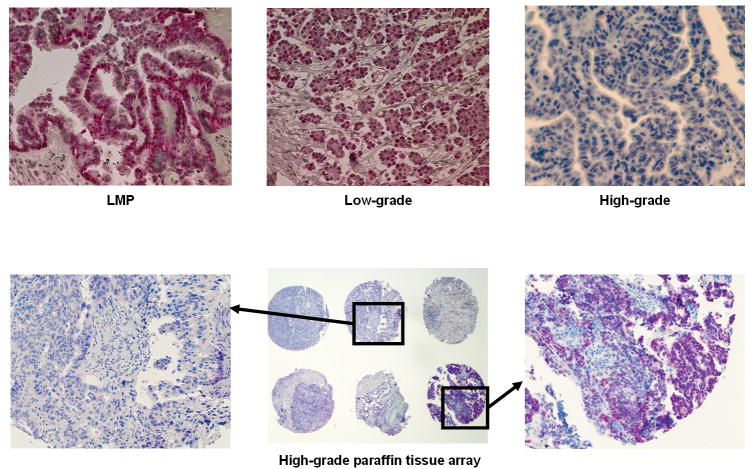
Figure 4.
A. Examples of PAX2 immunohistochemistry staining of individual paraffin sections from low-malignant potential tumors and low-grade and high-grade ovarian serous carcinomas (200× magnification) B. PAX2 immunohistochemistry staining of high-grade ovarian carcinoma paraffin tissue array sections. a, 200× magnification. b, 40× magnification. c, 200× magnification.

