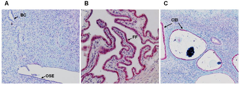
Figure 5.
PAX2 immunohistochemistry of A. normal human ovarian surface epithelial and benign ovarian cyst (BC) which have no PAX2 staining (100× magnification); B. fallopian tube fimbria (FF) demonstrating robust staining (200× magnification) and C. Ciliated epithelial inclusion (CEI) demonstrating robust staining (100× magnification).

