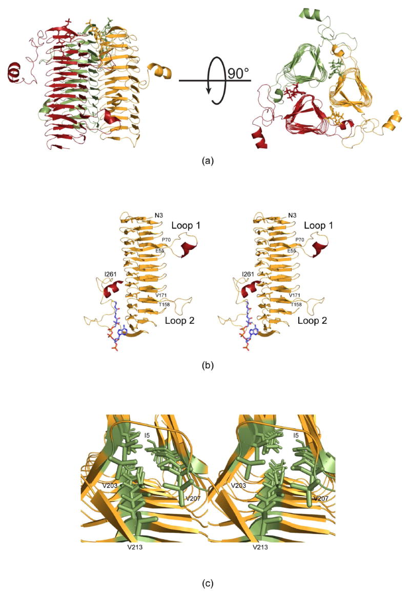
Figure 2.

Molecular architecture of QdtC. (a) Two views of the trimeric enzyme are shown with the individual subunits color-coded in green, red, and yellow. The polypeptide chains are depicted in ribbon representations whereas the acetyl-CoA ligands are shown as sticks. As can be seen, the three active sites of the trimer are located between subunits. (b) A stereo view of one subunit is displayed. The β-strands and α-helices are displayed in orange and red, respectively. (c) A close-up view is shown that highlights the three hydrophobic walls. These result from the hexapeptide sequence that dominates the primary structure of QdtC. All figures were prepared with the software package PyMOL (23).
