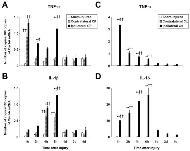
Figure 1.
Real-time RT-PCR analysis of temporal changes in choroidal (CP) and cortical (Cx) expression of TNF-α and IL-1β after TBI. The controlled cortical impact model of TBI in rats was used. (A, B) Changes in mRNA for TNF-α and IL-1β, respectively, in the ipsilateral and contralateral CPs, and in the CP from sham-injured rats (n=9–10 rats per time point). (C, D) Changes in mRNA for TNF-α and IL-1β, respectively, in the cerebral cortex (n=6 rats per time point). The number of copies of transcripts for each cytokine relative to the message for cyclophilin A (Cycl-A) is shown. Data represent mean values ± SEM. *P<0.05, **P<0.01 for the ipsilateral CP/Cx vs. contralateral CP/Cx (Newman-Keuls test). †P<0.05, ††P<0.01 for the ipsilateral or contralateral CP/Cx vs. the CP/Cx from sham-injured rats (Newman-Keuls test).

