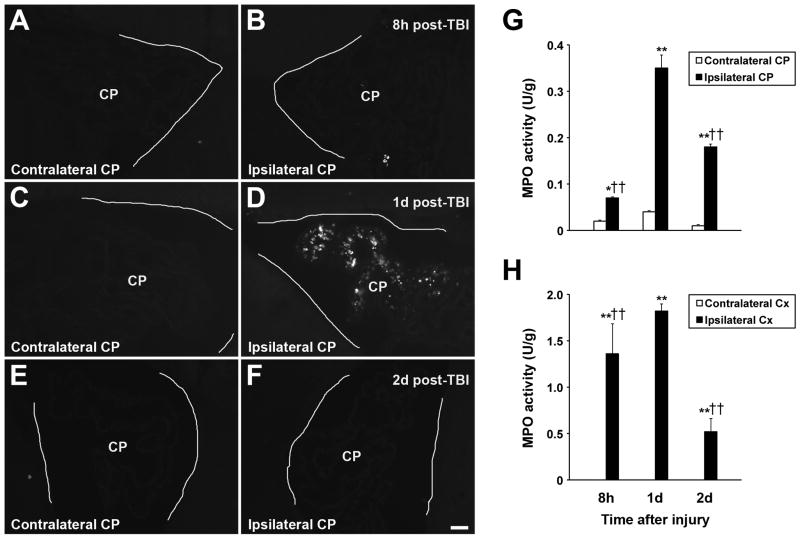
Figure 5.
Post-traumatic accumulation of neutrophils in the CP. (A, C, E) Low-power microphotographs of the contralateral CP at 8, 24, and 48 h post-TBI, respectively. (B, D, F) The ipsilateral CP at 8, 24, and 48 h post-TBI, respectively. The walls of the lateral ventricles are outlined. Neutrophils were detected by immunohistochemistry with anti-MPO antibody. Note that there was a significant delay between an increase in choroidal synthesis of chemokines and neutrophil accumulation in choroidal tissue. Neutrophils infiltrating the ipsilateral CP were found at 1 day after injury, whereas at 8 h post-TBI, these inflammatory cells were only sporadically observed to accumulate in the ipsilateral CP. This neutrophil influx was transient, as no neutrophils were found to infiltrate the ipsilateral CP at 2 days after TBI. Neutrophils did not accumulate in the contralateral CP. Bar: 50 μm. (G) The time course of post-traumatic changes in MPO activity in the CP (n=4 rats per time point). Low, but consistently detectable, MPO activity was found in the contralateral CP, which was likely associated with stromal macrophages and/or epiplexus cells normally present in the choroidal tissue. (H) Changes in MPO activity in the cerebral cortex adjacent to the lesion (n=4 rats per time point). Note that the peak levels (at 1 day post-TBI) of MPO activity in the injured cortex were ~5-fold higher than those observed in the ipsilateral CP. MPO activity was undetectable in the contralateral cortex. Data represent mean values ± SD. *P<0.05, **P<0.01 for the ipsilateral CP/Cx vs. contralateral CP/Cx (Newman-Keuls test). ††P<0.01 for the ipsilateral CP/Cx vs. the ipsilateral CP/Cx at 1 day post-TBI (Newman-Keuls test).

