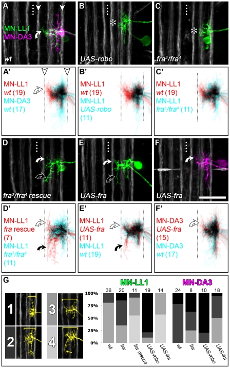
Figure 6. Robo and Frazzled mediate dendritic targeting in the intermediate and lateral neuropile.
(A–F) DiI/DiD-labellings of single MN-LL1 and MN-DA3 at 18.5 h AEL in fra- or robo-manipulated genetic backgrounds. (A′–F′) Cumulative plots generated from z-projections of various cells that were mapped onto a common reference grid using Fasciclin2-GFP-positive axon bundles as landmarks and shown in two channels, each representing one of two experimental conditions (n-numbers of cells in each plot are given in parentheses). Saturated colours indicate highly reproducible dendritic coverage at the respective relative position. In the wild-type, MN-LL1 can be distinguished from MN-DA3 by the presence of a dendritic subtree located in the intermediate neuropile (white curved arrow in [A, A′]). (B–C′) In fra mutants (C, C′) and when UAS-robo is expressed (B, B′; using CQ-GAL4) the intermediate dendrites of MN-LL1 fail to form (asterisks in [B and C]). (D, D′) Cell-specific expression of UAS-fra in the fra-mutant background rescues the intermediate MN-LL1 dendrites (white curved arrows) and frequently generates an ectopic posteriorly projecting intermediate branch (black curved arrows). (E, E′) Similarly, expression of UAS-fra in a wild-type background produces ectopic posteriorly projecting intermediate dendrites in MN-LL1 (black curved arrows). (F, F′) UAS-fra expression in MN-DA3 results in a subtle ectopic innervation of the intermediate neuropile (white curved arrows). (G) Illustration of and penetrance of four distinct medio-lateral dendritic morphologies for each motorneuron and genotype with indicated n-numbers above each bar. Dotted line: CNS midline. Scale bar 20 µm.

