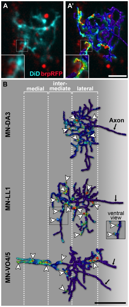
Figure 10. Medio-lateral distribution of excitatory presynaptic terminals on dendrites.
(A) Single confocal section of a motorneuron in freshly hatched larvae (21 h AEL) retrogradely labelled with DiD (cyan) with cholinergic presynaptic sites visualised with Cha-GAL4; UAS-brp-RFP (red). Part of the arbor (stippled outline) is shown enlarged in the inset in the bottom left-hand corner. (A′) The same confocal section is shown as in (A) and superimposed is a digital 3-D reconstruction of the entire dendritic arbor. Relative probabilities of synaptic connections were mapped onto the reconstructed arbour; colours towards the red spectrum indicating high probabilities based on brp-RFP fluorescence signal intensity and distance to dendrites (<400 nm). The insets in (A and A′) show an enlarged view of brp-RFP puncta and DiD-labelled dendrite in close apposition. (B) Dorsal views (a ventral view of part of the MN-LL1 arbor is shown in the inset) of representative reconstructed dendritic trees from dye-labelled MN-DA3, MN-LL1, and MN-VO4/5 with the distribution of putative synaptic sites mapped onto these as illustrated in (A and A′). Putative synaptic contacts (arrowheads) can be found on medial, intermediate, and lateral dendritic branches. Anterior is up. Scale bar: 5 µm in (A, A′) (2.5 µm for insets); 10 µm in (B).

