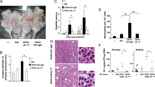
Fig. 5.
Effect of anti-IL-17 treatment of mice inoculated with VV in OVA-sensitized skin. (A) Primary lesions 8 days postinfection. (B) Size of the largest diameter of the primary lesion (n = 5–8 mice per group). (C) Number of satellite lesions (n = 5–8 mice per group). (D) Skin histology (400×, H&E stain). (E) Numbers of neutrophils in skin per 1,000 cells counted (n = 4 mice per group). (F) Viral load in internal organs. *, P < 0.05; **, P < 0.01.

