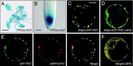
Fig. 3.
Tissue expression pattern and subcellular localization of AtTPST. (A) Histochemical staining of a 14-day-old Arabidopsis plant transformed with the AtTPSTpro:GUS gene. (Scale bar, 5 mm.) (B) Close-up of RAM in A. (Scale bar, 50 μm.) (C) Subcellular localization of the GFP-AtTPST fusion protein transiently expressed in Arabidopsis protoplasts. (Scale bar, 10 μm.) (D) Effect of BFA treatment (50 μg/mL) on GFP-AtTPST localization. (E) Colocalization of GFP-AtTPST with cis-Golgi marker, mRFP-SYP31. (F) Colocalization of GFP-AtTPST with mRFP-SYP31 after BFA treatment.

