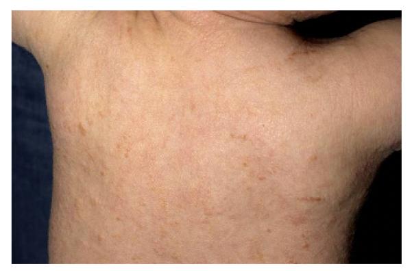
Figure 1. Diffuse Cutaneous Mastocytosis.

An infant with Diffuse Cutaneous Mastocytosis. The skin is diffusely infiltrated with mast cells demonstrating a peau d’ orange appearance without distinct uritcaria pigmentosa lesions.

An infant with Diffuse Cutaneous Mastocytosis. The skin is diffusely infiltrated with mast cells demonstrating a peau d’ orange appearance without distinct uritcaria pigmentosa lesions.