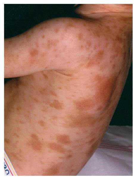
Figure 2. Urticaria Pigmentosa.

A child with Urtcaria Pigmentosa demonstrating typical lesions seen in patients with cutaneous mastocytosis. The lesions are varied in size and reddish-brown in color on a background of normal appearing skin.

A child with Urtcaria Pigmentosa demonstrating typical lesions seen in patients with cutaneous mastocytosis. The lesions are varied in size and reddish-brown in color on a background of normal appearing skin.