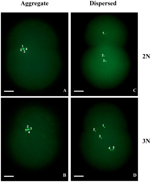
Figure 4. Sperm mitochondria segregation pattern in two-cell embryos.
Numbering was overlaid on the original photos to indicate how many sperm mitochondria were visible. In the “aggregate” pattern sperm mitochondria form an aggregate and stay in the same blastomere. In the “dispersed” pattern sperm mitochondria disperse randomly in the two blastomeres. A and B: Eggs from 98B (a male biased mother), sperm from 00WM2. C and D: Eggs from X102E (a female biased mother), sperm from Z101C. 2N: diploid, 3N: triploid.

