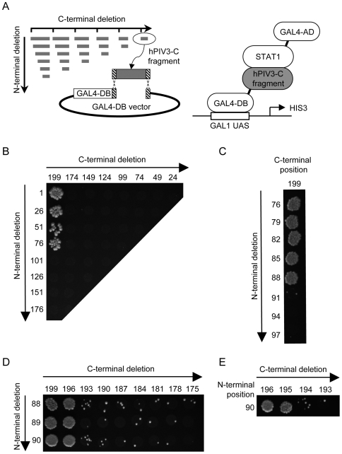
Figure 5. Systematic deletion-based mapping of a minimal hPIV3-C region interacting with STAT1.
(A) Fragments of hPIV3-C were generated by PCR using a matrix combination of specific primers (left panel), and introduced into Gal4-DB vector by gap-repair in yeast cells expressing STAT1 fused to Gal4-AD (right panel). Yeast cells were grown on selective medium lacking histidine and supplemented with 10 mM of 3-amino-triazole (3-AT) to test the interaction-dependent transactivation of HIS3 reporter gene. Vertical and horizontal axes indicate first and last AA residues of each fragment tested, respectively. (B), (C), (D) and (E) correspond to four iterations of this process. (E) The fourth led to the identification of a 106 AA encompassing position 90 to 195 of hPIV3-C as the minimal STAT1 binding domain.

