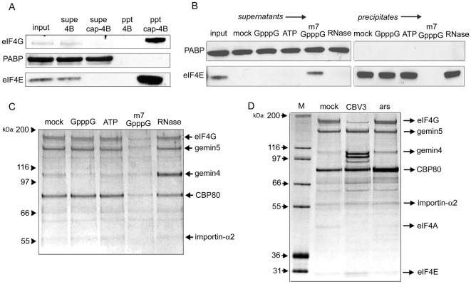
Figure 1. Characterization of proteins purified by cap-affinity chromatography.
(A) HeLa cytoplasmic lysate was applied to m7G-sepharose 4B or sepharose 4B alone. Precipitates (ppt) were washed extensively and then resuspended in sample buffer. Levels of eIF4G, PABP and eIF4E in input and supernatant (supe) samples were examined by western blot. Input samples represent 10% of total. (B) Cap-affinity chromatography was performed in the presence of the indicated nucleotide (0.1 mM final concentration) or 0.1 mg/ml RNase A and PABP/eIF4E were detected by western blot. (C) The same precipitate samples shown in (B) were subjected to silver stain detection. The identities of proteins determined by mass spectrometry are indicated. (D) HeLa lysates were derived from normally growing cells (mock), cells infected with CBV3 at four hours post-infection, or cells acutely stressed with 0.1 mM arsenite for 30 minutes. Proteins precipitated with cap-sepharose were then analyzed by silver stain.

