
Figure 4. mPar3 regulates asymmetric cell division of radial glial cells in situ.
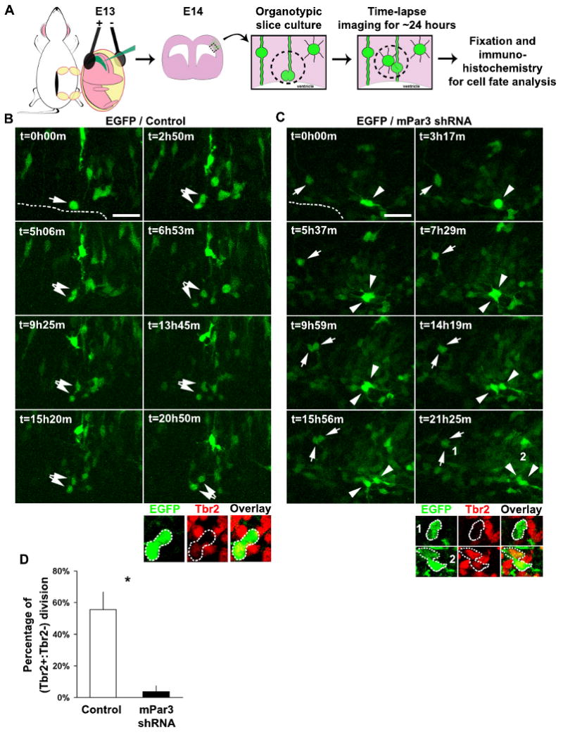
(A) Schematic representation of the procedure for examining the mode of division of radial glial cells in neocortical slices in situ. (B, C) Time-lapse images of radial glial cells expressing EGFP/Control shRNA (B) or EGFP/mPar3 shRNA (C) in organotypic cortical slice cultures. Arrows and arrowheads indicate dividing radial glial cells and their daughter cell pairs. Broken lines indicate the VZ surface. Immunohistochemistry analysis of EGFP-expressing (green) daughter cell pairs using the Tbr2 antibody (red) are shown at the bottom. Scale bars: 50 μm. (D) Quantification of the percentage of EGFP-expressing cells that divide asymmetrically to give rise to a Tbr2+ and a Tbr2- daughter cell (Control shRNA, 9 cells from three animals; mPar3 shRNA, 17 cells from eight animals). *, p<0.05.