
Some NLM-NCBI services and products are experiencing heavy traffic, which may affect performance and availability. We apologize for the inconvenience and appreciate your patience. For assistance, please contact our Help Desk at info@ncbi.nlm.nih.gov.

. Author manuscript; available in PMC: 2009 Sep 2.
Published in final edited form as: Circulation. 2009 Jan 26;119(5):709–716. doi: 10.1161/CIRCULATIONAHA.107.760819

Figure 2.

Figure 2

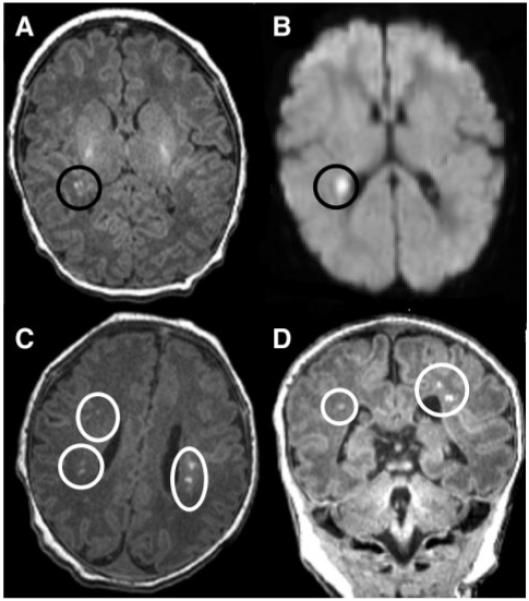
Brain MRI of preoperative infants with TGA: T1 imaging (A) and diffusion-weighted imaging (B) in a patient with mild PVL, which is a unifocal, small (<3 mm) white matter lesion. The lower MRI images demonstrate axial (C) and coronal (D) T1 imaging in a patient with bilateral, multifocal (moderate PVL) white matter disease.

HHS Vulnerability Disclosure