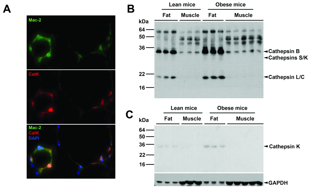
Figure 1. CatK expression in mouse adipose and muscle tissues.
A. Mouse visceral fat section co-immunostaining for macrophages (Mac-2) and CatK. Orange cells on the bottom panel were CatK-positive macrophages. B. Fat and muscle tissue lysate cathepsin active site [125I]-JPM labeling. Arrowheads indicate active cathepsins. C. CatK immunoblot analysis in mouse fat and muscle tissue extracts. Arrowhead indicates the 28-kDa mature CatK. Immunoblot for GAPDH was used for protein loading control.

