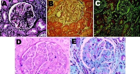
Figure 1.
Representative microphotographs of the diverse glomerular pathology seen in pSS. (A) Healed proliferative GN and focal global and segmental glomerulosclerosis (not shown). Healed proliferative glomerular lesions consistent with prior proliferative process (silver stain). (B) Minimal-change GN (hematoxylin and eosin). (C) Membranous nephropathy with localized subepithelial deposits confirmed on electron microscopy (not shown) (silver). (D) MPGN (hematoxylin and eosin). (E) Large subendothelial deposits characteristic of cryoglobulinemic GN (trichrome stain). Magnifications: ×100.

