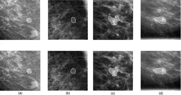
Figure 7.
Segmentation results for a benign lesion and a malignant lesion. The solid lines in the upper four images depict the lesion margin as outlined by a radiologist, and the solid lines in the bottom four images are segmentation results from our previously-reported automatic dual-stage method (Ref. 20). (a) CC view of a benign lesion, (b) the corresponding ML view of the benign lesion, (c) CC view of a malignant lesion, and (d) the corresponding ML view of the malignant lesion.

