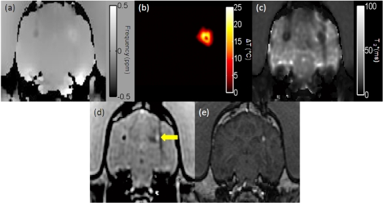
Figure 7.
The water PRF map of a canine brain used to create the temperature maps had a temperature sensitivity coefficient of −0.0098 ppm∕°C (a). The temperature map for the thermal therapy in the canine brain (b). The noise calculated in an unheated region contralateral to the therapy site was 0.34±0.09 °C. Using the CPD at , the uncertainty increased to a mean of 0.69±0.18 °C. A map generated by the SM algorithm (c). The T1-weighted images with correction simultaneously calculated at each acquisition along with the temperature maps (d). During treatment, a hyperintense area developed near the laser source, raising the suspicion of a treatment-induced hemorrhage (indicated by an arrow). The hyperintense lesion, clearly shown on (c), was confirmed as a treatment-induced hemorrhage on a post-treatment T1-weighted SPGR acquisition (e).

