Figure 4.
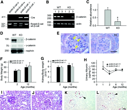
Mice with podocyte-specific deletion of β-catenin are healthy. (A) Genotyping of the mice by PCR analysis of genomic DNA. (B and C) RT-PCR analysis shows the glomerular β-catenin mRNA abundance in podo-β-cat−/− mice and their control littermates. WT, wild-type mice; KO, podo-β-cat−/− mice. Numbers (1, 2, and 3) denote each individual animal in a given group. *P < 0.05 versus WT (n = 3). (D) Western blot analysis of glomerular β-catenin protein in podo-β-cat−/− mice. Glomerular lysates were prepared from WT or KO mice and immunoblotted with antibodies against β-catenin, γ-catenin, and nephrin, respectively. (E) Immunohistochemical staining shows loss of β-catenin in glomerular podocytes. Arrowhead indicates positive staining for β-catenin in the glomeruli of WT mice at 1 d after ADR injection. No β-catenin staining was observed in the glomeruli of KO mice at the same conditions. (F through L) Mice with podocyte-specific ablation of β-catenin are phenotypically normal. There was little difference in (F) body weight, (G) kidney weight index, and (H) urinary albumin between KO mice and control littermates at different time points (n = 4 to 6). Kidney histology as shown by (I and J) periodic-acid-Schiff staining and (K and L) podocyte by WT-1 staining are normal in (J and L) KO mice, compared with (I and K) control littermates.

