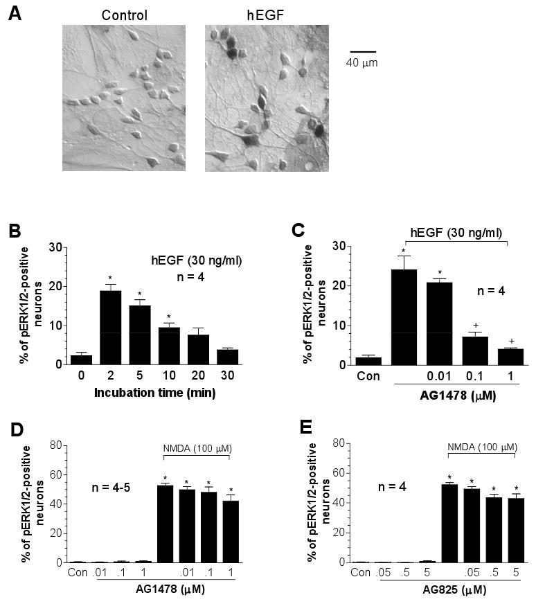
Fig. 2.

Effects of the receptor tyrosine kinase inhibitors on basal and NMDA-induced pERK1/2-immunoreactivity in cultured rat striatal neurons. (A) Immunocytochemical images illustrating increases in pERK1/2 neurons following hEGF incubation (30 ng/ml, 2 min). (B) Dynamic induction of pERK1/2 neurons following hEGF incubation (30 ng/ml, 2 to 30 min). (C-E) Effects of the EGF/ErbB1 inhibitor AG1478 or the ErbB2 inhibitor AG825 on hEGF- or NMDA-stimulated increases in the number of pERK1/2-positive neurons. The inhibitors were incubated 20 min prior to and during 2-min hEGF treatment or during 15-min NMDA treatment before fixation. Data are expressed in terms of the mean ± SEM of the percent change in numbers of the pERK1/2-positive neurons. *p < 0.05 vs. control (Con), and +p < 0.05 vs. hEGF alone (C).
