
Figure 7.
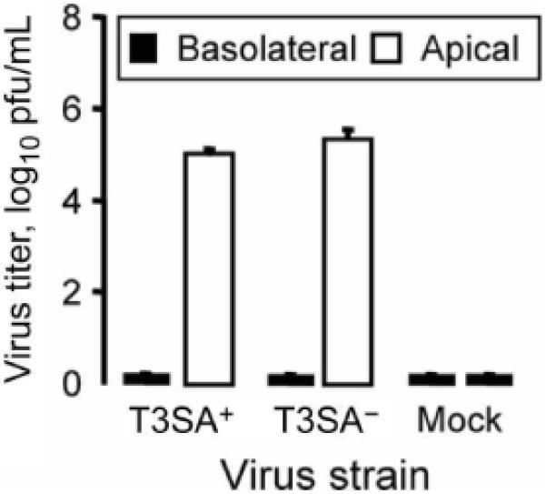
Reovirus shedding from infected airway epithelia. Epithelia were adsorbed basolaterally with T3SA+ (a monoreassortant reovirus strain that is capable of binding to sialic acid) (MOI, 200 pfu/cell) or T3SA- (a monoreassortant reovirus strain that is incapable of binding to sialic acid) (MOI, 50 pfu/cell), or they were mock treated. Washes of the apical surface or basolateral media were collected 21 days after adsorption, and viral titers were determined by plaque assay (n = 3). Error bars denote SDs.